OCT
Optical Coherence Tomography, or ‘OCT’, is a technique for obtaining sub-surface images of translucent or opaque materials at a resolution equivalent to a low-power microscope. It is effectively ‘optical ultrasound’. Imaging reflections presented in layers from within tissue to provide cross-sectional images enabling the doctor to see any abnormalities in the eye.
VISUAL FIELD
During a routine eye exam, Dr. Martin may want to determine through visual field testing the full horizontal and vertical range of what you are able to see peripherally. This range is commonly referred to as “side vision.”
Visual field tests assess the potential presence of blind spots (scotomas), which could indicate eye diseases and helps the doctor determine the presence of glaucoma, macular degeneration or your eye’s reactions to certain toxic medications. A blind spot in the field of vision can be linked to a variety of specific eye diseases, depending on the size and shape of the scotoma. Many eye and brain disorders can cause peripheral vision loss and visual field abnormalities.
OPTOMAP
The optomap ultra-wide field retinal image is a unique technology that captures more than 80% of your retina in one panoramic image while traditional imaging methods typically only show 15% of your retina at one time. The optomap retinal exam is fast, painless and comfortable. Nothing touches your eye at any time. It is suitable for the whole family. To have the exam, you simply look into the device one eye at a time (like looking through a keyhole) and you will see a comfortable flash of light to let you know the image of your retina has been taken.
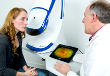 Under normal circumstances, dilation drops might not be necessary, but your eye care practioner will decide if your pupils need to be dilated depending on your conditions. The capture takes less than a second. Images are available immediately for review. You can see your own retina. You see exactly what Dr. Martin sees – even in a 3D animation.